Antibody selection guide
: To detect any target protein, there are more than one antibody to choose from. In order to narrow the choice of antibodies, select the appropriate antibody, you need to consider the following factors :? Type of analysis or application? Structural properties of the sample protein? Species of the sample Genus? Type of antibody host?
Antibody labeling and detection 1 Application type of analysis test ?? General antibody instructions list the type of analysis that the antibody has been verified by the test, such as: can be applied to WB? IHC? ICC? ELASA analysis, etc. If the antibody instruction manual The type of application not mentioned does not mean that the antibody is not suitable for this type of analytical application, but only that it has not been verified by this type of analytical test. If the antibody is not suitable for certain analytical tests, it will be marked on the antibody manual. It is not suitable for an analysis test.
2? The structural properties of the sample protein? Understanding the structural properties of the sample protein helps to select the most suitable antibody. At least two factors need to be considered (1). The domain of the sample protein to be tested: antibodies are composed of various immunogens It is prepared by immunizing the host. The immunogens include: full-length proteins, protein fragments, peptides, whole organisms (such as bacteria) or cells. The antibody instructions generally have a description of the immunogen. If the protein fragments or For a particular isoform or a certain region of the protein's full length, an antibody prepared with an immunogen containing this fragment domain must be selected. If you plan to use FACS to detect the surface protein of living cells, you need to select the extracellular domain containing the surface protein to immunize the prepared antibody.
(2) Sample extraction or processing process: Some antibodies require samples to undergo some special processing, for example: many antibodies only recognize reduced and denatured protein samples whose epitopes have been exposed and are not hindered by secondary and quaternary structures. In some aspects, some antibodies only recognize proteins in their natural folded state. When selecting antibodies for immunohistochemistry, it should be noted that some antibodies only recognize unfixed frozen tissues, while others are suitable for formaldehyde-fixed paraffin-embedded tissues that do not require antigen repair, delinking, and cross-linking. The application part is marked on the antibody manual
3 Species of the sample?
Antibodies of the same species or cross-reactivity should be selected. Antibodies may cross-react with the same target protein of different species. Because of the high amino acid sequence homology, if the type of the sample is not included in the cross-reactive species on the antibody manual In the table, it does not mean that the antibody is not suitable for detecting the protein of the species, but only that the species has not been tested and verified by this antibody. The cross-reaction should be predicted by sequence alignment. Expasy and NCBI BLAST can be used Perform protein homology comparison of different species.
4 Selection of primary antibody host species
In general, when using a conjugated secondary antibody in combination with a primary antibody without conjugate, the species selection of the primary antibody host animal is more important. For immunohistochemistry, try to select a primary antibody of a different germline species from the sample, In order to avoid cross-reaction between the secondary antibody and the endogenous immunoglobulin in the sample, for example: detecting mouse sample protein, you should not choose a mouse or rat-derived primary antibody, preferably a rabbit-derived primary antibody, the secondary antibody Then select anti-rabbit IgG coupled with detection molecules (enzyme, fluorescein, biotin, etc.). If the primary antibody with conjugate is selected, the above situation is not applicable. Except for immunohistochemistry, other detection methods for samples without endogenous immunoglobulin will have little effect on the host species of the antibody, such as IgG-free Western blotting detection of cell lysate samples, however, tissue lysates containing serum and tissue culture supernatants contain immunoglobulins, reduced denatured samples contain IgG, IgG molecules 50 and 25 are combined in Western blot detection kDa heavy and light chain bands.
5 Selection of secondary antibodies
The secondary antibody should be selected from the same species as the primary antibody used. For example, if your primary antibody is a mouse monoclonal antibody, the secondary antibody should be anti-mouse secondary. It is recommended to check the secondary antibody instructions to ensure that the antibody is suitable for your detection application. The secondary antibody is generally connected to fluorescein FITC or luminophore. 6 Selection of double-stained antibodies ?? Double immunostaining of cell cultures or tissue sections with unconjugated primary antibodies requires that the primary antibodies come from different species and that the secondary antibodies recognize one of them, and the secondary antibody instructions should describe it and other species Is the source of immunoglobulin cross-adsorbed? 7Fluorescent and chemiluminescent labels? The label attached to the secondary antibody is used to detect the binding of the antibody. The selection of the label depends on several parameters: detection method: fluorescence or colored precipitation, the fluorescent label is emitted when excited by light of a specific wavelength Visible light, the table below lists the molecular characteristics of several fluoresceins: fluorescein color excitation wave emission molecular weight Aminomethylcoumarin (AMCA) Blue 353 440 410 Fluorescein (FITC) Green 495 528 390 Fluorescein (DTAF) Green 495 528 530 Rhodamine (TRITC) Red 550 570 444 Texas Redtm Red 596 620 625 Cy2tm Green 489 505 897Cy3tm Orange 552 565 949Cy3.5tm Scarlet 581 596 1,286Cy5tm Far-Red 650 667 975Cy5.5tm Near Infra-Red 678 703 1,312Cy7tm Near Infra-Red 743Co ) Red 488 575 240,000B-Phycoerythrin (BPE) Red 545 575 240,000C-Phycocyanin Red 618 640 110,000R-Phycocyanin Red 615 620 110,000 secondary antibody ligation labeling enzymes HRP and AP and its substrate produce a colored precipitate horseradish peroxidation Horseradish peroxidase (HRP) Alkaline phosphatase (AP) AEC (red) Fast red (pink) DAB (brown) INT (yellow / brown)? NBT (brown / purple)? New Fuchsin (red)? TNBT (purple)? Vega red (pink) mounting medium (immunohistochemistry only). AEC, Fast Red, INT or other water-based light-emitting groups are alcohol-soluble and require water-based sealing. Light-emitting groups other than the above are organic, so it is best to use organic sealing media. Fluorescein labels require an aqueous mounting medium, phycoerythrin (phycocyanin / phycoerythrin requires an aqueous mounting medium that does not contain glycerin, because of its effect of quenching fluorescence, biotinylated antibodies are usually used to enhance affinity Heparin-biotin-enzyme or fluorescein complex (ABC reagent or avidin or streptavidin ligase or fluorescein signal)
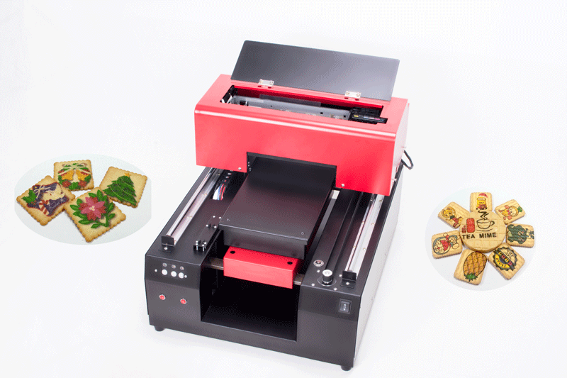
A4 size edible printing printer/Biscuit Printer with edible ink can print on all kinds of food such as Macaron, M&M candies, Chocolate, cake, Biscuit,macaron etc .Each item can be printed as a unique one with colorful photoes . or you can print one color ink according with your request.

5-35º
Name
Biscuit printer
Print head
DX5
Print size
21*38cm
Print color
CMYK+LC LM
Print speed
A4 photo/163s
Printing resolution
5760*1440dpi
Nozzles
90*6=540
Interface
USB 2.0
Net weight/Gross weight
35kg/42kg
Printer size
57*48*44cm
Height adjustment
Handle/Automatic
Working power
110-220 50-60Hz 30W
Operation system
Windows 2000/XP/WIN7/Vista etc.
Temperature
Biscuit Printer
Biscuit Printer,Cookies Printer,Automatic Biscuit Printer,Digital Biscuit Printer
Shenzhen Refinecolor Technology Co., LTD. , https://www.rfcprinter.com