Peking University develops a new generation of micro microscopes for real-time brain analysis

In the new millennium, brain science research has become a hot spot. If a worker wants to do something good, he must first sharpen his tools. To better explore the human brain, you must have better instruments and tools. At present, a core direction of national brain science programs is to create research tools for panoramic analysis of brain-connected maps and functional dynamic maps. Among them, how to break the scale barrier and integrate microscopic neuron and synaptic activity with the brain's overall activity and individual behavior information is a key challenge in the field.
Recently, Nature Magazine published a research report from China on this aspect. This paper mainly shows the research results of "Ultra-high-space-resolved miniaturized two-photon in-vivo microscopic imaging system" - a new generation of high-speed high-resolution miniaturized two-photon fluorescence microscope, and acquired the mouse in the process of free behavior. Clear, stable images of brain neurons and synapses.
The research results were derived from the national major scientific research equipment development project organized by the National Natural Science Foundation of China, and nine projects were selected at that time. One of them was the "super-high-space-resolved miniaturized two-photon in-vivo microscopic imaging system" led by Academician Cheng Heping of Peking University. At that time, it also received funding support of 72 million yuan.
In the past three years, the Institute of Molecular Medicine, the School of Information Science and Technology, the Dynamic Imaging Center, the School of Life Sciences, and the School of Engineering of the Peking University, together with the Interdisciplinary Team of the Chinese Academy of Military Medical Sciences, completed this research and development work. The group successfully developed a new generation of high-speed high-resolution miniaturized two-photon fluorescence microscope, and obtained clear and stable images of brain neurons and synapses in mice during free behavior. The research paper was submitted in December 2016 and was officially released on May 29, 2017 in the Nature Journal.
According to the information provided by the official, the product has better optical tomography and deeper biological tissue penetration than single photon excitation. Its lateral resolution reaches 0.65μm, and the imaging quality can reach commercial large-scale desktop two-photon fluorescence. The microscope level is superior to the miniaturized wide field microscope developed by the United States. The microscope uses a two-axis symmetric high-speed MEMS rotating mirror scanning technology, the imaging frame rate has reached 40Hz (256 * 256 pixels), and has multi-area random scanning and 10,000 lines per second line scanning capability.
In addition, a self-designed photonic crystal fiber that conducts a 920 nm femtosecond laser is the first to realize the effective use of a fluorescent probe (such as GCaMP6), the most widely used indicator neuron activity in the field of brain science, by miniature two-photon microscopy.
At the same time, the use of a flexible fiber bundle for the reception of fluorescent signals solves the problem that the activity and behavior of the animal are disturbed due to the dragging of the fluorescent transmission cable. In the future, the combination of optogenetics technology is expected to accurately manipulate the activities of neurons and neural circuits while imaging structurally and functionally.
It is worth mentioning that the microscope weighs only 2.2 grams and can record the dynamic signals of dozens of neurons and thousands of synapses in real time on the cranial window of the small animal's head. On large animals, it is expected to achieve more Long-term observation of different brain regions of the probe wearing and multiple cranial windows.
The reason why this research is significant is mainly because it provides important high-end instruments for the research of brain science and artificial intelligence. Specifically, micro-two-photon fluorescence microscopy has changed the way cells and subcellular structures are observed in free-living animals, and can be used in natural behaviors such as foraging, breastfeeding, jumping, fighting, play, and sleep. Or long-term observation of multi-scale, multi-level dynamic changes such as synapses, neurons, neural networks, and remotely connected brain regions before, during, and after study.
In fact, imaging technology has always been the main driving force behind the advancement of life sciences. Historically, X-ray, holography, CT computed tomography, electron microscopy, MRI nuclear resonance imaging, and ultra-high resolution microscopy have all contributed to the advancement of science and technology and have also won the Nobel Award.
Before today's press conference, the results were reported at the end of 2016 at the American Neuroscience Annual Meeting and the May 2017 Cold Spring Harbor Asian Brain Science Conference, and were received by domestic and foreign neuroscientists including many Nobel Prize winners. Recognition. Professor Alcino J Silva, president of the Asian Brain Science Conference at Cold Spring Harbor, and the famous American neuroscientist at the University of California, Los Angeles, believes that "this microscope will change the way we observe cells and subcellular structures in free-living animals." System Neurobiology Entering a new era, through the observation of complex biological events of identifiable cells and subcellular structures in cell populations, to gain a deeper understanding of the core engineering principles of evolutionary brain loops to achieve complex behaviors ""
At the same time as the successful development of this technology, the team also established a company called “Super Vista†and obtained financing from the Synergy Innovation Fund and Xike Angel. The company will be supported by Peking University under the premise of meeting the policy of Peking University. Commercialization promotion. The team's next focus is still on technology iterations and new product development.
Children Furniture Sets Desk Chairs
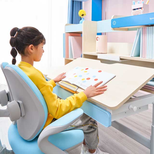
igrow study table and chair, the height of the table and chair can be adjusted according to the height of the child. In addition to the most basic "cushion height" that can be adjusted, the "armrest and back cushions" can also be adjusted according to the child's thigh length and waist height. Adjust so that the child does not sit in the wrong position due to the improper height of the table and chair. And the chair is designed according to children's ergonomics, unlike ordinary chairs that are straight up and down, there is a double-back curve design, which supports the child's back and ensures that the waist and hips fit perfectly with the chair.
Through multiple adjustable ergonomic designs and healthy and environmentally friendly materials, Aigole provides children with long-lasting healthy companionship and learning assistance.

Kids Wooden Table Chairs,Combo Desk And Chair,Kids Table With Storage,Adjustable Kids Table
Igrow Technology Co.,LTD , https://www.szigrowdesks.com